Crown hair transplant · Macclesfield
Restore the crown. Protect the future.
A crown—or vertex—hair transplant moves selected donor follicles into thinning areas around the natural whorl. Because the crown can consume many grafts while native hair continues to thin, diagnosis, direction and lifetime donor planning matter as much as the operation itself.
An assessment may lead to medical treatment first, FUE or FUT surgery, a staged plan, observation—or a recommendation not to transplant the crown.
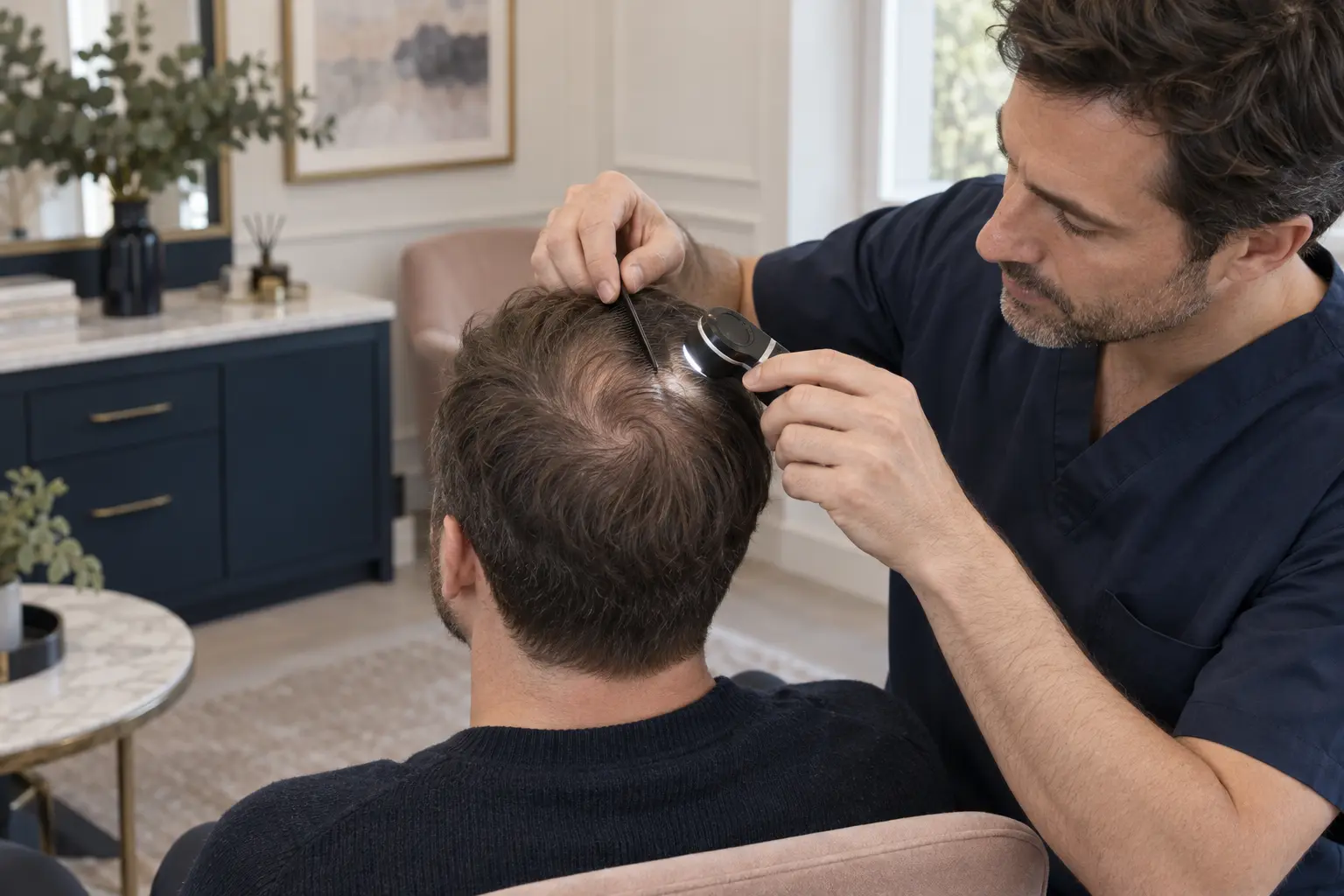
Read the pattern first
Crown thinning is a pattern—not a circle to fill.
The vertex curves away from view and hair radiates from a whorl. A convincing result depends on how the central pivot, surrounding miniaturisation and future loss relate to each other.
Visible scalp is influenced by the size of the thinning area, hair calibre, curl, colour contrast, density and the angle at which hair leaves the scalp. A graft number alone cannot predict coverage.
The whorl centre
The pivot may sit centrally or off-centre, with clockwise or anticlockwise flow. Recipient sites should respect the patient’s own pattern.
Design: locate the pivotThe transition ring
Miniaturised hairs around the visible centre can make the true treatment field larger than the obvious patch seen in a photograph.
Diagnosis: map the edgeDiffuse vertex loss
Hair may be present but progressively finer across a broad area. Surgery through unstable native hair can increase shock-loss and planning risk.
Stability: measure changeThe donor relationship
Every crown graft comes from the same finite donor reserve that may later be needed for the hairline, mid-scalp or repair work.
Strategy: protect supplyA crown quote without a whole-scalp plan is incomplete.
The apparent gap is only one part of the decision. Donor capacity and the likely future pattern must be examined before grafts are committed.
The procedure, made clear
A crown transplant redistributes follicles. It does not create new supply.
Follicular units are harvested from an assessed donor area by FUE or FUT and placed into surgeon-created recipient sites around the vertex. The visual effect comes from survival, calibre, direction, layering and contrast—not from recreating original density.
Transplanted follicles generally retain characteristics of their donor origin, but growth is not guaranteed and the non-transplanted hair remains biologically vulnerable. Poorly timed crown work can leave an isolated “island” or ring as surrounding hair recedes.
Candidate selection
Enough stability. Enough donor. A realistic coverage goal.
Crown surgery is considered in the context of age, diagnosis, rate of change, frontal and mid-scalp priorities, scalp health, donor quality, hair characteristics and willingness to protect native hair where clinically appropriate.
A crown transplant may be considered when…
Surgery may be unsuitable or premature when…
The assessment pathway
Plan from the whorl out—and from the future back.
A credible consultation records the crown you have, the pattern it may become and the donor reserve required to keep the whole scalp coherent over time.
Confirm the diagnosis
We review onset, progression, family history, shedding, symptoms, illness, medicines, previous treatment and whether further investigation is indicated.
Map the visible and hidden field
Standardised photographs and examination define the whorl, transition zone, diffuse miniaturisation and relationship to the mid-scalp.
Examine under magnification
Hair calibre variation, follicular density, scalp condition and the presence of miniaturised native hair help judge stability and shock-loss risk.
Measure the donor
Density, calibre, follicular-unit composition, laxity where relevant, miniaturisation and previous scarring inform safe harvest options.
Build a lifetime graft budget
The likely needs of the hairline, mid-scalp, crown and any future repair are weighed before allocating a finite number of donor follicles.
Agree the sequence
Medical management, observation, FUE, FUT, staging, combined approaches and no surgery are compared with expected coverage, risks and cost.
Two harvest routes
FUE and FUT change the donor scar—not the need for crown design.
Both methods can provide follicular-unit grafts for the vertex. The method should follow donor anatomy, hairstyle, previous surgery, graft requirement, scarring preference and long-term harvest strategy.
Important: “DHI” usually describes a way of placing grafts, not a third source of donor follicles. Whatever device is used, diagnosis, safe harvesting and surgeon-led recipient-site design remain essential.
Follicular unit excision
Follicular units are removed individually through small circular donor wounds and then placed into crown recipient sites.
Follicular unit transplantation
A donor strip is removed and dissected into follicular units before the donor wound is closed.
Three linked design zones
The eye reads flow before it counts follicles.
Whorl pivot
The natural centre is identified rather than automatically redrawn. Small errors here can make every surrounding angle look artificial.
Radial flow
Recipient-site direction and angle change continuously around the pivot to overlap coverage without creating upright or crossing hairs.
Transition perimeter
The outer edge blends into miniaturised native hair and the mid-scalp. A hard circular boundary can become conspicuous as loss progresses.
If surgery is appropriate
Harvest conservatively. Rebuild the spiral deliberately.
The exact surgical sequence varies by method and patient. Your consent should identify the operating surgeon, each team member’s role, the proposed graft range, donor limits, risks, aftercare and what happens if growth is less than expected.
Reconfirm the plan
Diagnosis, medicines, photographs, donor boundaries, crown design, staged priorities and consent are checked before treatment begins.
Prepare and anaesthetise
The donor and recipient areas are prepared and local anaesthetic is administered. Any sedation arrangement should be individual and explained in advance.
Harvest the grafts
Follicular units are removed by the agreed FUE or FUT approach while donor distribution, transection and future reserve are monitored.
Inspect and protect
Grafts are examined, organised by characteristics and kept appropriately hydrated while the recipient design is created.
Create the crown pattern
Recipient sites follow the changing angle and direction of the native whorl, using distribution to create coverage without a hard border.
Place, check and discharge
Grafts are placed atraumatically, the donor and crown are inspected, and written care, medicines, contacts and reviews are provided.
Healing and growth
The grafts settle first. The visual result takes months.
Your own surgical team’s written instructions take priority. These phases are broad guideposts, not deadlines; FUE and FUT donor healing, shedding and crown growth vary.
0–2
Early protection
Redness, tenderness, pinpoint crusting and minor oozing can occur. Swelling may develop away from the crown. Avoid pressure, rubbing and unapproved products.
3–7
Careful cleansing
Crusts remain visible while donor sites or the FUT closure heal. Washing, sprays, sleep position and any dressing follow the clinic’s instructions.
1–2
Surface healing
Crusting usually settles progressively. Non-dissolvable FUT closure material, if used, is reviewed according to the surgeon’s schedule.
2–8
Shedding phase
Transplanted shafts commonly shed. Temporary shock loss can also affect native or donor hair, particularly where miniaturisation is present.
3–6
Early growth
New hairs may begin appearing at different times and can initially be fine or uneven. Early photographs do not predict final density.
10–18
Crown maturation
Calibre, length and visual coverage may continue to improve over an extended period. Final review also considers ongoing native loss.
Understand the possible complications.
Hair transplantation is surgery. Crown-specific planning reduces avoidable problems but cannot guarantee graft survival, density or satisfaction.
Contact the surgical team promptly for severe or increasing pain, active bleeding, marked swelling, spreading redness, discharge, fever, breathing difficulty, visual symptoms or any unexpected deterioration.
Plan beyond the first procedure
Transplanted follicles may persist. The surrounding crown can still recede.
A durable strategy protects the native hair where clinically appropriate, preserves donor options and reviews the whole pattern rather than chasing every new patch with surgery.
Consider stabilisation first
Evidence-based medical treatment may be discussed after individual assessment, contraindication review and shared decision-making.
Prioritise visible framing
The frontal scalp often delivers greater visual impact. Crown allocation should not compromise a coherent long-term hairline and mid-scalp plan.
Reserve donor capacity
A conservative first procedure can leave options for progression, repairs or refinement rather than exhausting the safe donor area.
Review with comparable images
Consistent lighting, hair length and angles help distinguish genuine growth from styling, wetness, contrast or camera effects.
Your clinical team
Know who diagnoses, designs and operates.
Crown transplantation combines medical diagnosis with irreversible donor harvesting. Your plan should state who is responsible for assessment, extraction, recipient-site creation, graft placement and postoperative review.
Dr Fida Ul Haq
Hair-loss assessment and long-term planning
Dr M Muhammad
Lead hair transplant surgeon
Before you decide: verify the clinician’s current registration and role, ask who performs every surgical stage, and take time to consider the diagnosis, alternatives, graft budget, costs, recovery and material risks without pressure.
Questions worth asking
Clear answers before donor hair is committed.
These are general explanations, not personal medical advice. Suitability and a responsible graft range require an in-person clinical assessment.
What is a crown hair transplant?
A crown hair transplant moves follicular-unit grafts from an assessed donor area into thinning areas at the vertex. Recipient sites are designed around the natural whorl so the transplanted hair follows changing radial angles and blends with remaining native hair.
Is a crown hair transplant permanent?
Transplanted follicles generally retain characteristics of their donor origin, but no clinic can guarantee that every graft will grow or remain unchanged. The surrounding non-transplanted hair can continue to thin, so “permanent cure” is not an accurate description.
Who may be suitable for crown transplantation?
Potential candidates usually have a confirmed transplantable diagnosis, reasonably stable loss, a healthy scalp, adequate stable donor hair and realistic coverage expectations. Rapid progression, active scalp disease, diffuse donor miniaturisation or higher frontal priorities may make surgery premature or unsuitable.
How many grafts does the crown need?
There is no responsible universal number. The area, existing miniaturised hair, whorl pattern, hair calibre, colour contrast, desired visual change, donor reserve and needs elsewhere on the scalp all affect the proposed range. Photographs alone are not enough for a final quote.
Is FUE or FUT better for the crown?
Neither method is automatically better for the crown. Both can supply follicular units; they differ mainly in donor harvesting and scarring. The choice depends on donor anatomy, graft requirement, hairstyle, previous surgery, healing preference and the long-term harvest plan.
Does a crown hair transplant leave scars?
Yes. FUE leaves many small distributed extraction scars, while FUT leaves a linear donor scar. Recipient sites also heal with microscopic scarring. Visibility varies with technique, healing, hair length, contrast and whether the donor is overharvested.
When will crown transplant results be visible?
Transplanted shafts commonly shed in the first weeks. New growth may begin after several months and then gain length and calibre gradually. Crown maturation can continue into the 10-to-18-month range, but timing, survival and density vary between patients.
How much does a crown hair transplant cost?
Cost depends on the diagnosis, surgical method, proposed graft range, complexity, operating team, facility, aftercare and whether treatment is staged. A written quote should explain what is included, possible additional costs, finance terms and the clinic’s approach if growth is below expectation.
A consultation, not a commitment
Plan the whole scalp before spending donor on the crown.
Meet the clinical team in Macclesfield for diagnosis, magnified crown and donor assessment, whorl mapping, treatment comparison and a realistic long-term graft strategy.
This page provides general information and is not a diagnosis, personalised treatment recommendation or guarantee of outcome.